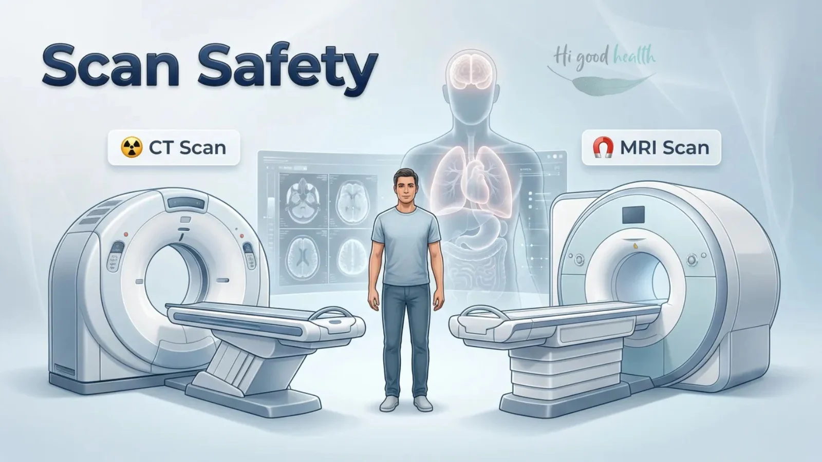
Ever felt uneasy when a doctor asks for a “scan”? Words like “radiation” and “dye” can sound worrying, and it is natural to wonder whether these tests are safe.
At a Glance: Is MRI Dangerous, and Which Scans Actually Use Radiation?
MRI is not dangerous in the way most people fear it is. It works with a strong magnet and radio waves and emits no ionising radiation at all. Neither does ultrasound. The scans that do use radiation are X-ray, CT, mammography, DEXA, fluoroscopy, PET and nuclear medicine studies.
The doses are small, and the unit is unfamiliar, which is most of where the worry comes from. A chest X-ray is about 0.1 mSv — roughly ten days of the radiation you receive anyway from the ground, the sun, your food and the air.
A CT of the abdomen is about 10 mSv, which is a few years of it. Those two numbers put the whole question in proportion.
The useful question is therefore not how much radiation but will this scan change what happens next. And for MRI specifically, the safety conversation is not about radiation at all — it is about metal: pacemakers, certain implants, and anything ferromagnetic near a very strong magnet.
Routine blood tests do not always answer every health question. To understand more about advanced diagnostic tests, read Hidden Blood Tests for Peak Health (That Your Insurance Won’t Cover), Part 1 of 3.
Let’s clear the confusion and look at the facts about medical scans, radiation, and those so-called liquids, so you can make informed decisions about your health.
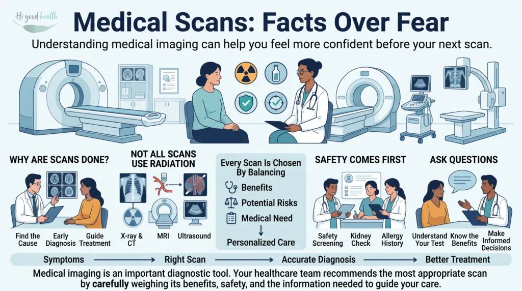
The Imaging Arsenal: Which Scans Use Radiation, and Which Do Not
Medical imaging is like a team of different specialists, each with its own strengths. From finding a tiny fracture to spotting complex diseases, these tests play a major role in diagnosis and treatment.
- X-ray (Radiography): The classic, quick, and affordable test for bones, lungs, and things like kidney stones. It uses a small amount of radiation.
- CT Scan (Computed Tomography): Think of it as a highly detailed 3D X-ray. It takes multiple X-ray slices to build cross-sectional images of bones, organs, and blood vessels. It is very useful in emergencies, cancer staging, and complex diagnoses.
- MRI (Magnetic Resonance Imaging): The soft-tissue expert. MRI uses strong magnetic fields and radiofrequency waves, and it does not expose patients to ionising radiation. MRI is especially useful for the brain, spinal cord, joints, muscles, and other soft tissues.
- Ultrasound (Sonography): Sound waves do the work here. This radiation-free scan uses high-frequency sound to create real-time moving images. It is commonly used for pregnancy scans, checking organs such as the liver and kidneys, and assessing blood flow.
- Endoscopy: Not an external scan, but an important internal examination. A flexible tube with a camera is inserted into the body, such as through the mouth for gastroscopy or through the rectum for colonoscopy, to see and sometimes treat internal problems. No radiation is involved.
- PET Scan (Positron Emission Tomography): PET scans use a small amount of radioactive tracer, most commonly fluorodeoxyglucose (FDG), a glucose analogue, to measure activity in tissues. Areas with higher glucose use, such as many cancers or sites of inflammation, appear brighter on the scan.
- Mammography: A special X-ray test for breast tissue, important for early detection of breast cancer.
- DEXA Scan (Bone Densitometry): A low-dose X-ray test that measures bone mineral density, important for diagnosing osteoporosis.
- Fluoroscopy: A real-time imaging method that uses continuous or pulsed X-rays to create moving images of internal structures. It is often used to guide minimally invasive procedures, such as catheter placement, joint injections, and angiography, and to study movement in organs, including swallowing and the digestive tract.
- Nuclear Medicine Scans: A broad category, such as bone scans or thyroid scans, in which a very small amount of radioactive material helps show how an organ is functioning.
Understanding thyroid health starts with knowing which tests matter. To know more about thyroid testing, read Hidden Blood Tests for Peak Health: The Complete Guide.
- Angiography: Uses X-rays and a special dye to map blood vessels and find blockages or abnormalities.
- Elastography: A modern technique, often combined with ultrasound or MRI, that measures tissue stiffness, which can be useful in conditions such as liver fibrosis.
How Much Radiation Is in a Scan? The Dose Facts
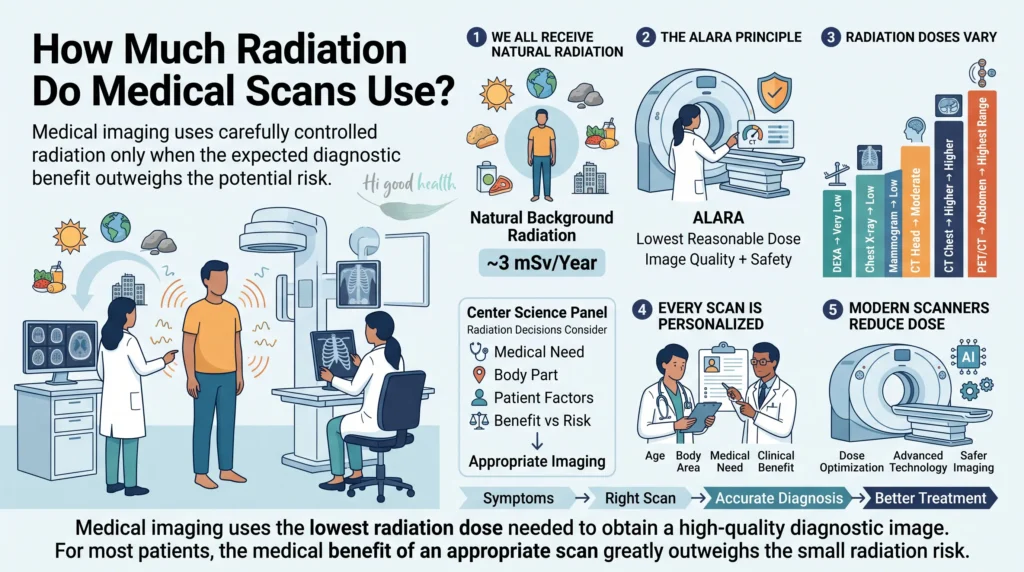
The word “radiation” can sound alarming, but medical scans use very small, controlled doses. We are all exposed to natural background radiation every day, from the earth, the sun, building materials, food, and radon gas.
Worldwide it averages about 2.4 mSv per person per year. Most of India sits below that, at roughly 1 to 2 mSv, because indoor radon levels are generally lower than in colder countries with sealed housing.
India also has one of the most-studied high-background regions in the world. The monazite sands along coastal Kerala and parts of Tamil Nadu deliver several times the national average to people who have lived there for generations, and the large cohort studies run in those districts have not found the excess cancer rate that a simple dose-equals-risk model would predict.
That does not make radiation harmless. It does mean the doses involved in ordinary medical imaging sit in a range where the risk is small and hard to measure, which is the honest way to describe it.
There is no fixed “maximum number of scans” a person can have. Instead, medical professionals follow the ALARA principle: As Low As Reasonably Achievable. This means using the smallest possible dose needed to get a clear, diagnostic image.
The risk from radiation adds up over a lifetime and depends on the type of scan, the area scanned, age, the organs exposed, the dose from each examination, and the underlying medical condition.
Here is a simplified look at typical radiation doses, measured in millisieverts (mSv), compared with a natural background of about 3 mSv per year. That is a deliberately conservative benchmark, higher than the Indian average, so for most readers here the equivalent times in the last column are if anything understated:
Scan Type | Approximate Effective Dose (mSv) | Equivalent Natural Background Time | Key Takeaway |
|---|---|---|---|
Chest X-ray | 0.1 | ~10 days | Very low, often the first step. |
Mammogram | 0.4 | ~2 months | Low dose for important breast screening. |
CT Head | 2 | ~8 months | Moderate, but often critical for neurological issues. |
CT Chest | 7 | ~3 years | Higher, but provides valuable detail for lungs and chest. |
CT Abdomen/Pelvis | 10 | ~3–4 years | Among the higher doses, used for detailed abdominal and pelvic conditions. |
PET/CT (Whole Body) | 10–25 (tracer + CT) | ~4–10 years | Combines functional and anatomical imaging, often used for cancer staging. |
DEXA Scan | 0.001 | ~0.5 days | Extremely low, used for bone density. |
Important Note: The estimated increase in lifetime cancer risk from a single CT scan, such as 10 mSv, is tiny — roughly 0.05%. For context, the lifetime cancer risk from all causes is about 40–50%. In most cases, the diagnostic benefits outweigh this very small risk.
The values above are approximate effective doses for common examinations. Actual exposure varies depending on the scanner, imaging protocol, body size, and clinical need. Many modern CT systems use dose-reduction technology that can lower radiation exposure while maintaining image quality.
Contrast Dye: What It Is, and Who Needs to Be Careful
You are right to ask about the “dye.” Many scans, especially CT and MRI, use contrast agents to improve visibility of organs, blood vessels, or abnormalities. This liquid is usually swallowed, injected into a vein, or sometimes given rectally.
Types of contrast agents:
- Iodine-based contrast: Commonly used for CT scans and X-ray procedures like angiography. It contains iodine, which absorbs X-rays and makes blood vessels and organs easier to see.
- Gadolinium-based contrast: Used for MRI scans. Gadolinium changes the magnetic properties of water molecules and improves MRI signal in certain tissues.
- Barium-based contrast: Often a chalky liquid swallowed for X-ray or fluoroscopy studies of the digestive tract, such as a barium swallow. It coats the digestive lining so it can be seen clearly.
Are they harmful?
Although contrast agents are generally safe and leave the body fairly quickly, they can sometimes cause side effects. Serious reactions are rare, but it is important to inform your medical team about:
- Allergies, especially to iodine or previous contrast agents, or a history of severe allergies or asthma. Reactions can range from mild hives or itching to severe breathing difficulty or anaphylaxis.
- Kidney problems. Kidney function is often checked before iodine-based and gadolinium-based contrast. Poor kidney function can make it harder for the body to clear the contrast, which may cause complications. With gadolinium, there is a very rare but serious condition called Nephrogenic Systemic Fibrosis (NSF) in patients with severe kidney disease.
- Asthma, heart conditions, and diabetes. These conditions may increase the risk of certain reactions.
- Medicines. Some medicines, such as certain diabetes drugs like metformin, may need to be adjusted before or after a contrast scan.
Your medical team will usually screen you carefully for risks before giving contrast.
Is MRI Dangerous? Why MRI and Ultrasound Use No Radiation at All
MRI and ultrasound do not use ionising radiation, but they still have their own safety points to keep in mind.
- MRI concerns: Powerful magnets are the main issue. Metallic implants such as pacemakers, certain clips, or shrapnel can be affected. Always tell your doctor and MRI staff about any metal in or on your body. Many modern implants, including some pacemakers, joint replacements, vascular stents, and surgical clips, are MRI-compatible under specific conditions, but each implant must be checked carefully before the scan.
- Claustrophobia: The enclosed space of an MRI machine can be difficult for some people.
- Loud noise: Ear protection is important during the scan.
- Contrast agents: Gadolinium-related concerns are discussed in the section above.
- Ultrasound concerns: Diagnostic ultrasound is generally considered extremely safe. It uses very low power levels, so risks such as tissue heating or cavitation are minimal and mostly apply to much higher-power therapeutic ultrasound.
- For fetal ultrasound, it is safe when used for medical reasons, but non-medical “keepsake” ultrasounds are generally discouraged, following the ALARA principle.
X-Ray and CT in Pregnancy: What the Guidance Actually Says
Pregnancy does not automatically mean imaging cannot be done. If a scan is medically necessary, the healthcare team will choose the safest option that still gives the needed information.
- Ultrasound is usually the first choice during pregnancy because it uses sound waves and does not involve ionising radiation.
- MRI is also considered safe during pregnancy when clinically needed, as it uses magnetic fields and radiofrequency waves instead of X-rays.
- Gadolinium-based MRI contrast is generally avoided during pregnancy unless the benefit clearly outweighs the possible risk to the baby.
Imaging tests that use ionising radiation, such as X-rays, CT scans, fluoroscopy, and nuclear medicine scans, are not automatically unsafe during pregnancy.
- The risk depends on the radiation dose, the stage of pregnancy, and the area being scanned.
- Most routine diagnostic scans expose the baby to radiation levels well below the range associated with harm, especially when the abdomen or pelvis is not directly imaged.
- When medically necessary, for example in trauma, suspected pulmonary embolism, appendicitis, or stroke, the benefit of timely diagnosis usually outweighs the small risk.
Imaging decisions during pregnancy should always be made individually through discussion between the patient and the healthcare team.
Guidelines for Diagnostic Imaging During Pregnancy and Lactation
Is MRI Safer Than CT? How X-Ray, CT and MRI Compare
On radiation alone, yes: MRI wins, because it has none. But that is not the comparison a doctor is making, and it is not the one you should make either. The three tests answer different questions, and picking the one with the lowest dose is only sensible if it can actually see what needs to be seen.
| X-ray | CT | MRI |
|---|---|---|---|
Ionising radiation | Yes, very low | Yes, the highest of routine imaging | None |
Best at seeing | Bone, lungs, stones, hardware | Bleeds, trauma, lungs, abdomen, cancer staging | Brain, spinal cord, joints, ligaments, soft tissue |
Time in the machine | Seconds | Under a minute for most studies | 20 to 60 minutes, and noisy |
Main safety issue | Cumulative dose over many films | Dose, especially in children and repeat scanning | Metal. Pacemakers, some implants, metal fragments |
Typical cost in India | Lowest | Middle | Highest, and often the longest wait |
Where the substitution genuinely works: for a suspected ligament or cartilage injury, a soft-tissue mass, or most brain and spine questions that are not an emergency, MRI is both the better test and the zero-dose one. For a child with recurrent abdominal pain, an ultrasound first is standard practice for exactly this reason.
Where it does not: in acute trauma or a suspected bleed in the brain, CT is faster and answers the question that has to be answered in minutes. Refusing a CT there to avoid 2 mSv trades a small, statistical, long-term risk for a large, immediate one.
It is reasonable to ask whether a scan is necessary. It is not reasonable to decline the one the emergency actually calls for.
The one thing worth carrying with you: keep your own record of scans, with dates and body regions. Radiology departments in India do not share a common patient dose registry, so if you move between hospitals nobody is adding it up.
A note on your phone is enough, and it lets the next doctor make a better decision than a guess.
Choosing a Safer Imaging Centre in India
Healthcare technology keeps improving, and new machines are designed to reduce radiation while improving image quality. Here is how to identify centres that focus on patient safety:
Not every health concern needs another blood test. See Are Frequent Blood and Other Diagnostic Tests Really Necessary? for the non-invasive ways to monitor your body.
- Look for accreditation: Prefer facilities accredited by recognised organisations such as the American College of Radiology (ACR), or equivalent accredited imaging standards where available.
- Ask about dose-reduction technology: When booking a CT scan, ask whether they use Iterative Reconstruction (IR) or Automatic Exposure Control (AEC). These technologies can significantly lower the radiation dose without affecting image clarity.
- Ask about child-focused safety practices: If the scan is for a child, ask whether the centre follows Image Gently® principles. For adults, look for Image Wisely® practices.
- Discuss with your doctor: Always ask why the scan is needed and whether a lower-radiation alternative, such as MRI or ultrasound, could provide similar information.
PET-CT and PET-MRI: Higher Doses, Specific Reasons
These are advanced hybrid scans that combine functional imaging (PET) with anatomical imaging, giving doctors the best of both worlds.
- PET (Positron Emission Tomography): The functional part. A radioactive tracer shows metabolic activity, meaning how cells are working. This is useful for finding active areas such as cancer cells.
- CT (Computed Tomography): The anatomical part of PET-CT. It provides sharp images of bones and organs and helps pinpoint where the PET activity is located.
- MRI (Magnetic Resonance Imaging): The anatomical part of PET-MRI. It offers better soft tissue detail without radiation, helping doctors locate PET activity more precisely in areas like the brain or pelvis.
In simple terms:
Feature | PET-CT | PET-MRI |
|---|---|---|
Combines | PET (Function) + CT (Anatomy) | PET (Function) + MRI (Anatomy) |
Strength | Widely available, excellent for whole-body cancer staging, strong anatomical localisation | Lower radiation dose, better soft tissue detail, useful for brain, pelvis, and paediatric imaging |
Primary Use | Common for cancer detection, staging, and monitoring | Used more often for specific cancers, neurological studies, and selected cases |
Radiation | Yes, from the PET tracer and CT component | Yes, from the PET tracer only; MRI itself has no radiation |
Availability | More widely available | Less common, more specialised, and usually more expensive |
The choice between these tests depends on the clinical question and the patient’s condition. The medical team will select the best option.
Key Takeaways
- MRI and ultrasound emit no ionising radiation. If radiation is your worry, these two are not the tests to worry about.
- X-ray, CT, mammography, DEXA, fluoroscopy, PET and nuclear medicine scans do use radiation — in doses ranging from about a tenth of a millisievert to around twenty-five.
- A chest X-ray is about ten days of natural background radiation. A CT of the abdomen is a few years of it. Both are small numbers; only one is worth thinking twice about.
- MRI risk is about metal, not radiation. Tell the centre about any pacemaker, implant, clip, coil or metal fragment before the appointment, not on the day.
- There is no lifetime cap on scans. Doctors work to ALARA — as low as reasonably achievable — weighing the dose against what the scan will change.
- Ultrasound and MRI are the preferred tests in pregnancy, and a single X-ray away from the abdomen is not a reason to panic. Tell the radiographer you are pregnant, or might be.
- Keep your own list of scans with dates and body regions. No one in India is adding them up for you.
- The question to ask is not “how much radiation” but “what will you do differently depending on the result”. A scan that changes nothing is the only scan whose dose was definitely not worth it.
Frequently Asked Questions
Do CT scans and X-rays cause cancer?
Diagnostic scans use very low radiation doses. The estimated lifetime cancer risk from a single CT scan is tiny, and the medical benefits of diagnosis usually outweigh the small risk.
What is the difference between a CT scan and an MRI?
CT uses X-rays to show bones and dense organs quickly, which makes it useful in emergencies. MRI uses magnets without radiation to show fine details in soft tissues like the brain and joints.
Is contrast dye used in scans safe?
Contrast agents are generally safe, but they can cause mild side effects such as nausea. Serious reactions are rare, but you should inform your doctor about kidney problems, asthma, or iodine allergies.
How many CT scans are safe in a lifetime?
There is no fixed maximum number. Doctors follow the ALARA principle and use the lowest dose that still gives a useful result.
Can I have an MRI if I have metal implants?
MRI magnets can affect metals such as pacemakers or shrapnel. Many modern implants are safe under specific conditions, but you must tell the staff about any metal in your body.
Are medical scans safe for children?
Yes, but children are more sensitive to radiation. Look for facilities that follow child-focused low-dose practices such as Image Gently.
Does radiation stay in my body after a scan?
For standard X-rays and CT scans, the radiation passes through immediately and does not remain in the body. PET and nuclear medicine scans use radioactive tracers that stay only briefly until the body clears them.
Why do doctors sometimes order multiple scans?
Different scans show different things. A doctor may order a CT for bone detail and an MRI for soft tissue to get a complete picture and confirm the diagnosis.
Is MRI dangerous?
Not in the way most people mean. An MRI scanner uses a strong magnetic field and radio waves, and produces no ionising radiation at all, so there is no radiation dose to add up and no cumulative risk from having had several. The genuine cautions are different ones: metal. A pacemaker, an implanted defibrillator, some older aneurysm clips, cochlear implants and metal fragments in the eye can be unsafe near a scanner, which is why the centre asks about them in detail. Most modern orthopaedic implants, stents and dental work are fine, but the radiology team must be told what you have before the appointment rather than on the day. Beyond metal, MRI is loud, it takes twenty minutes to an hour, and the tunnel is uncomfortable if you are claustrophobic, all of which can be planned around.
How much radiation is in a CT scan compared with an X-ray?
Roughly twenty to a hundred times more, depending on which CT and which X-ray. A chest X-ray delivers about 0.1 mSv, which is close to ten days of the natural background radiation you receive anyway. A CT of the head is around 2 mSv, a CT of the chest around 7 mSv, and a CT of the abdomen and pelvis around 10 mSv, or several years of background. A PET-CT, which combines both, is higher again at roughly 25 mSv. Those figures are averages and vary with the machine, the protocol and your body size; a modern scanner running a low-dose protocol can be well below them.
Is it safe to have an X-ray during pregnancy?
Tell the radiographer you are pregnant, or think you might be, before anything is switched on. That single step is what the safety system depends on. Where imaging is needed in pregnancy, ultrasound and MRI are preferred because neither uses ionising radiation. Where an X-ray or CT is genuinely necessary, it is usually still done, with shielding and the lowest workable dose, because an undiagnosed serious condition in the mother is the larger risk. A single X-ray of a limb, chest or teeth delivers a very small dose to the uterus and is not a reason for alarm. What should always be a conversation with your doctor rather than a decision made alone is a CT of the abdomen or pelvis.
How many scans are too many? Is there a lifetime limit?
There is no fixed number, and any article that gives you one is inventing it. Radiology works to a principle called ALARA, as low as reasonably achievable: each scan is justified on its own merits, at the lowest dose that still answers the question. Risk accumulates over a lifetime and depends on your age, which organs were in the beam and how large each dose was, so ten dental X-rays and ten abdominal CTs are not remotely the same thing. The practical safeguard is not a counter but a question: ask what the scan will change. If the answer is that treatment will be the same either way, that is the scan worth questioning. Keep your own dated list of scans, because no hospital in India is keeping it for you.
Glossary
- ALARA: “As Low As Reasonably Achievable”; a safety principle used to keep radiation exposure as low as possible while still getting a useful image.
- Angiography: An imaging test that uses X-rays and contrast dye to view blood vessels and detect blockages or abnormalities.
- Contrast agent / contrast dye: A substance given by mouth, injection, or rectum to make organs, blood vessels, or tissues easier to see on scans.
- CT scan: Computed Tomography; a scan that uses X-rays to create detailed cross-sectional images of the body.
- DEXA scan: Dual-energy X-ray absorptiometry; a low-dose X-ray test used to measure bone mineral density and diagnose osteoporosis.
- Effective dose (mSv): A measure of radiation exposure used to estimate the possible risk from an imaging test.
- Endoscopy: A procedure that uses a flexible tube with a camera to look inside the body; it does not use radiation.
- FDG: Fluorodeoxyglucose; a radioactive tracer commonly used in PET scans to measure metabolic activity.
- Fluoroscopy: A real-time X-ray imaging method used to guide procedures and observe movement inside the body.
- Gadolinium-based contrast: A contrast agent used in MRI to improve the visibility of certain tissues and structures.
- Ionising radiation: High-energy radiation that can change atoms and is used in X-rays, CT, fluoroscopy, and some nuclear medicine scans.
- MRI: Magnetic Resonance Imaging; a scan that uses magnetic fields and radiofrequency waves, not ionising radiation, to image soft tissues.
- Nuclear medicine scan: A scan that uses a small amount of radioactive material to show how organs or tissues are functioning.
- PET scan: Positron Emission Tomography; an imaging test that uses a radioactive tracer to show metabolic activity in the body.
- Radiation dose: The amount of radiation received during an imaging test, often measured in millisieverts (mSv).
- Ultrasound: An imaging test that uses sound waves to create images and does not use ionising radiation.
- X-ray: A quick imaging test that uses a small amount of radiation to view bones and some organs.
References
- RadiologyInfo.org — MRI Safety
Radiological Society of North America (RSNA) & American College of Radiology (ACR) - American Institute of Ultrasound in Medicine (AIUM) — Official Safety Statements on Diagnostic Ultrasound
AIUM Safety Statement - American College of Obstetricians and Gynecologists (ACOG) — Guidelines for Diagnostic Imaging During Pregnancy and Lactation
ACOG Committee Opinion - American College of Radiology (ACR) — ACR Accredited Facility Search Tool
ACR Accreditation Search - Image Gently® Alliance — Radiation Protection in Paediatric Medical Imaging
Image Gently - Image Wisely® — Radiation Safety in Adult Medical Imaging
Image Wisely
Disclaimer
The content provided on higoodhealth.com is for general informational purposes only. While we strive to offer accurate and up-to-date content, much of the information is derived from publicly available sources and personal research. We do not make any warranties about the completeness, reliability, or accuracy of this information.